Brazilian researchers last month presented a new face for a 9,000-year-old skull discovered in 1953 near the West Bank city of Jericho.
The man’s skull, partially covered in plaster and with shells for the eyes, was one of seven discovered by archaeologist Kathleen Kenyon. All are believed to have served as part of a Neolithic ritual related to ancestor worship.
In 2016, the British Museum used micro-computed tomography, or micro-CT, to scan the skull and the product 3D-printed reproduction of the original skull and reconstruction of the person’s face for an exhibit. Other skulls are on display in Britain, Canada and Jordan, and at the Rockefeller Archaeological Museum in East Jerusalem.
In December, the Brazilian team published A new detailed 3D face, with artistically rendered head hair and facial hair.
To create a new face, the researchers used anatomical deformation and statistical inference based on micro-CT scans – techniques used to plan plastic surgery and create prosthetics.
Cicero Moraes, the Brazilian graphics expert who led the project, told the Live Science website that the method provides “greater anatomical, anatomical and statistical coherence”.
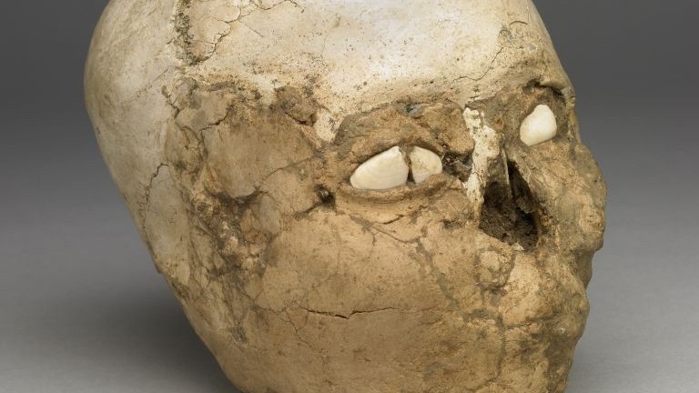
A 9,000-year-old Neolithic plaster skull from the collection of the British Museum. (courtesy: british museum)
“I wouldn’t say ours is an update, it’s just a different approach,” to the British Museum’s model, he said.
Moraes worked with Thiago Beini, a dental surgeon and assistant professor at Brazil’s Federal University of Uberlândia, and Moaquir Elias Santos, an archaeologist at the Museum of Archeology in Ponta Grossa.
Archaeologists have determined that the man died in his 30s or 40s because of the way the wound on the skull had healed.
Moraes said the skull is unusual because its upper part is larger than average.
The head, like others found in the area, appears to have been artificially stretched, possibly due to the man being tightly bound during puberty. The reason for this practice is unknown.
Also last month, Egyptian and British scientists Used CT software to create new detailed images of ancient ruler Ramses II.
The scientists first used CT scans of the pharaoh’s mummy and applied analysis software to parse the details. The experts were then able to differentiate between the skull and other materials used during the engraftment process and create 3D images of the skull. The face was then reconstructed using Egyptian measurements of the folds of the facial muscles.
Times of Israel staff contributed to this report.